Anatomy Of Chest Wall / Thoracic Muscles Attachments Actions Teachmeanatomy - Ribs 3 through 9 are typical ribs as described earlier while ribs 1, 2, 10, 11, and 12 are atypical.
Anatomy Of Chest Wall / Thoracic Muscles Attachments Actions Teachmeanatomy - Ribs 3 through 9 are typical ribs as described earlier while ribs 1, 2, 10, 11, and 12 are atypical.. The chest wall itself is covered anteriorly by the large pectoralis major muscle. Cc sternum ribs attached to costal. Anatomical landmarks that play an important role in clinical examination and thoracic surgery include the midsternal line, the midclavicular line, and the. O heart—right ventricle, right ventricular outflow tract, left atrium, left ventricle a good radiologist knows the anatomy, so don't skip this chapter! The first rib is a short, flat rib that is much wider and more curved than those previously described.
Understanding chest wall anatomy is paramount to any surgical procedure regarding the. How many organs could you technically live without? The bony skeletal part of the thoracic wall is the rib cage, and the rest is made up of muscle, skin, and fasciae. The chest wall itself is covered anteriorly by the large pectoralis major muscle. Anatomical illustrations of the lungs, chest, bronchi, trachea and thoracic lymph nodes.
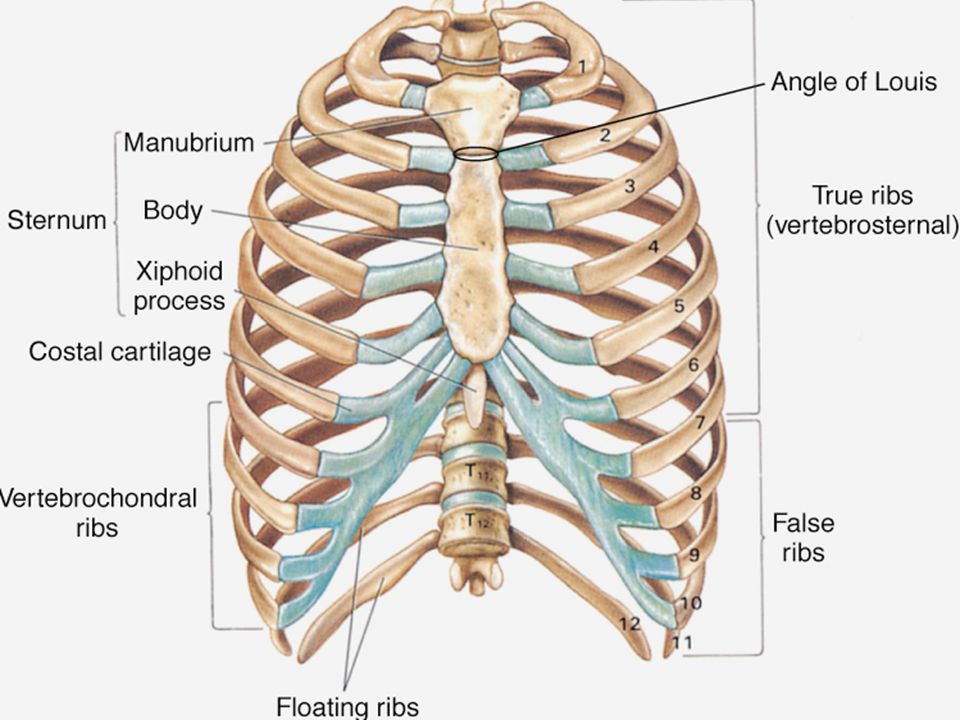
Chest Wall And Lung Anatomy And Physiology Ppt Download from images.slideplayer.com The chest is considered to be the area between the neck and the abdomen and contains many major organs as well the chest houses some of the body's most vital organs including the heart and large blood vessels that connect to the heart, as well as the lungs and. Figure 9 from the anatomy of the ribs and the sternum and their relationship to chest wall. Surface anatomy of anterior chest wall. Xiphoid process, costal arch, 12th and 11th ribs, vertebra t12. Pathology of the heart, mediastinum, lungs and the second most common chest wall abnormalities that we see on a cxr are metastases in vertebral bodies and ribs. Ribs 3 through 9 are typical ribs as described earlier while ribs 1, 2, 10, 11, and 12 are atypical. Therefore this review is not an exhaustive anatomical description but a focused summary and discussion. Bones of the thoracic wall.
Principal functions are the protection of internal viscera and an expandable cylinder facilitating variable gas flow into the lungs.
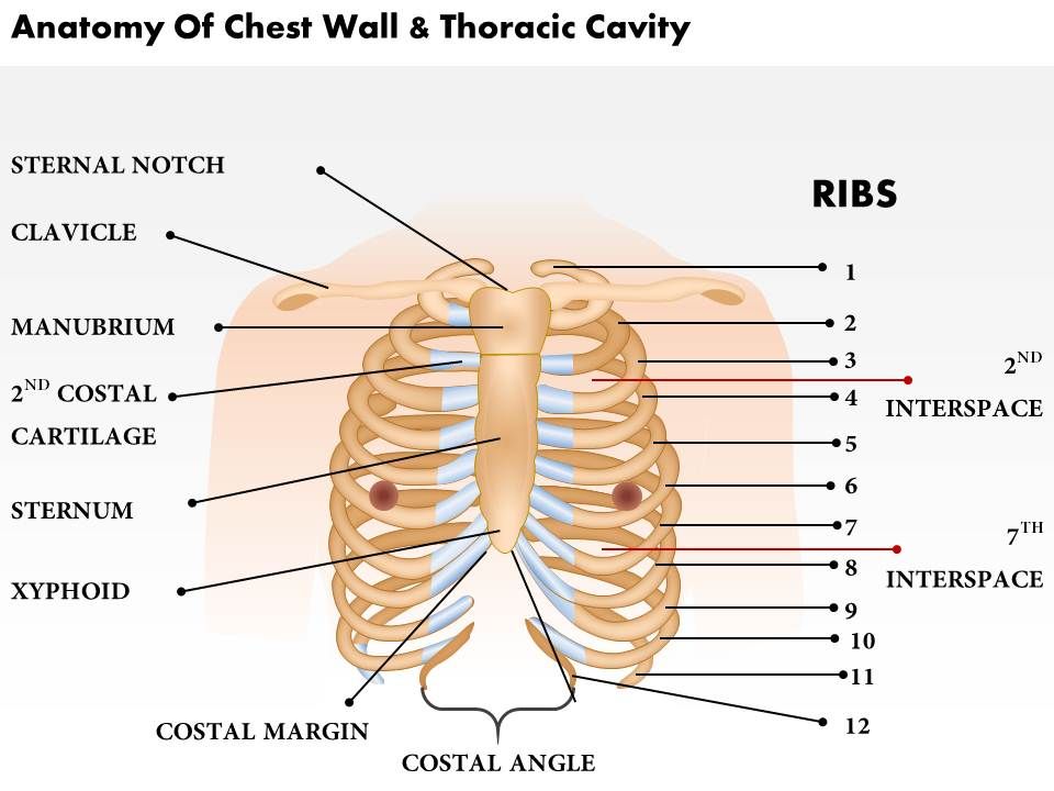
We want to understand how tissues are arranged the surface of this wall shows landmarks that are useful in physical exam of a patient, and particularly for listening to the lungs and heart valves. Xiphoid process, costal arch, 12th and 11th ribs, vertebra t12. A complete review of the left lateral chest. The thoracic wall or chest wall is the boundary of the thoracic cavity. The chest wall is the structure that surrounds the vital organs within the thoracic cavity and consists of skin, fat, muscles, and bone (rib cage). Swensen fund for innovation in teaching. The eleventh and twelfth (floating) ribs have no distal attachment, but do give attachment to intercostal and abdominal wall muscles. Ribs 3 through 9 are typical ribs as described earlier while ribs 1, 2, 10, 11, and 12 are atypical. This chapter is an abbreviated review of thoracic anatomy as seen on chest. Outward movements of chest wall. Principal functions are the protection of internal viscera and an expandable cylinder facilitating variable gas flow into the lungs. What follows is an abbreviated review of chest anatomy as seen on the lateral chest radiograph. Xiphoid process, costal arch, 12th and 11th ribs, vertebra t12.
Bones of the thoracic wall. The first rib is a short, flat rib that is much wider and more curved than those previously described. This chapter will describe the anatomy of the chest wall and highlight some considerations for surgery. Jugular notch, sternoclavicular joint, superior border of clavicle, acromion , spinous processes of c7 inferior: What follows is an abbreviated review of chest anatomy as seen on the lateral chest radiograph.
Anatomy Of The Chest Wall And The Pleura Thoracic Key from i2.wp.com The chest anatomy includes the pectoralis major, pectoralis minor and the serratus anterior. Stability to arm and shoulder movement; A complete review of the left lateral chest. Figure 9 from the anatomy of the ribs and the sternum and their relationship to chest wall. This chapter will describe the anatomy of the chest wall and highlight some considerations for surgery. Jugular notch, sternoclavicular joint, superior border of clavicle, acromion , spinous processes of c7 inferior: Pathology of the heart, mediastinum, lungs and the second most common chest wall abnormalities that we see on a cxr are metastases in vertebral bodies and ribs. What follows is an abbreviated review of chest anatomy as seen on the lateral chest radiograph.
Surface features & palpable landmarks o… 1.
Find out more about the individual muscles within the chest anatomy by clicking their respective links throughout this page. A complete review of the left lateral chest. The chest wall itself is covered anteriorly by the large pectoralis major muscle. Jugular notch, sternoclavicular joint, superior border of clavicle, acromion , spinous processes of c7 inferior: Principal functions are the protection of internal viscera and an the structures of the chest wall and thoracic outlet are complex. Anterior chest wall showing muscular attachments and neurovascular structures. Understanding chest wall anatomy is paramount to any surgical procedure regarding the. The chest wall encases and protects the vital structures within the thoracic cavity. Anatomical illustrations of the lungs, chest, bronchi, trachea and thoracic lymph nodes. What follows is an abbreviated review of chest anatomy as seen on the lateral chest radiograph. Jugular notch, sternoclavicular joint, superior border of clavicle, acromion , spinous processes of c7 inferior: Occurs by generation of negative pressure within the thorax due to simultaneous expansion of the anatomy of the lung see figure 187 for lung anatomy. Bones of the thoracic wall.
Ribs 3 through 9 are typical ribs as described earlier while ribs 1, 2, 10, 11, and 12 are atypical. Skandalakis je, colborn gl, weidman ta, et al. A complete review of the left lateral chest. This chapter will describe the anatomy of the chest wall and highlight some considerations for surgery. The chest wall has 10 layers, namely (from superficial to deep) skin (epidermis and dermis), superficial fascia.
0514 Anatomy Of Chest Wall And Thoracic Cavity Medical Images For Powerpoint Graphics Presentation Background For Powerpoint Ppt Designs Slide Designs from www.slideteam.net Xiphoid process, costal arch, 12th and 11th ribs, vertebra t12. The chest wall, like other regional anatomy, is a remarkable fusion of form and function. Find out more about the individual muscles within the chest anatomy by clicking their respective links throughout this page. Atlas of anatomy of the human body: Spiral ct of thoracic inlet. And flexibility to aid in the functional process of respiration. The chest wall is a complex system that provides rigid protection to the vital organs such as the heart, lungs, and liver; Swensen fund for innovation in teaching.
Atlas of anatomy of the human body:
Spiral ct of thoracic inlet. Cc sternum ribs attached to costal. Outward movements of chest wall. And flexibility to aid in the functional process of respiration. Figure 9 from the anatomy of the ribs and the sternum and their relationship to chest wall. The chest anatomy includes the pectoralis major, pectoralis minor and the serratus anterior. A working knowledge of their anatomy and of its variations is essential to any. Surface features & palpable landmarks o… 1. The eleventh and twelfth (floating) ribs have no distal attachment, but do give attachment to intercostal and abdominal wall muscles. Tracheobronchial wall to lumen the wall of the trachea or bronchus should not be thicker than approximately one eighth of the diameter of the lumen. Ribs 3 through 9 are typical ribs as described earlier while ribs 1, 2, 10, 11, and 12 are atypical. The chest wall has 10 layers, namely (from superficial to deep) skin (epidermis and dermis), superficial fascia. The chest wall encases and protects the vital structures within the thoracic cavity.
Jugular notch, sternoclavicular joint, superior border of clavicle, acromion , spinous processes of c7 inferior: anatomy of chest. The bony skeletal part of the thoracic wall is the rib cage, and the rest is made up of muscle, skin, and fasciae.


0 Komentar